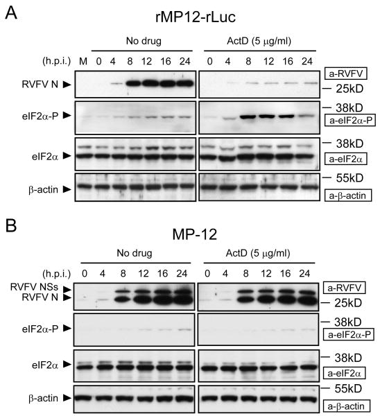
Figure 2.
Status of eIF2α phosphorylation in rMP12-rLuc-infected cells and MP-12-infected cells in the presence of transcriptional inhibitors. The Fig is adapted from Ikegami et al.[26] VeroE6 cells were mock infected (M) or infected with rMP12-rLuc (A) or MP-12 (B) at an moi of 3, and then immediately treated with ActD or left untreated (No drug). Samples were harvested at the indicated time points post infection for Western blot analysis. RVFV N protein, NSs proteins, phosphorylated eIF2α, total eIF2α, and β-actin are shown by arrowheads. The data are representative of three independent experiments.

