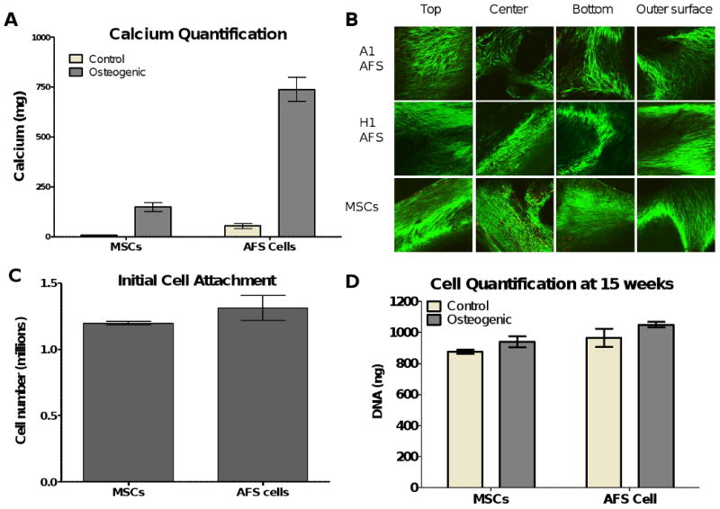
Figure 4.

A. The calcium produced by the cells during the 15 weeks in culture was compared and quantified using the Arsenazo III reagent. 25% of the scaffold was used for this comparison. The AFS cells produced significantly more mineralized matrix than the MSCs when cultured in osteogenic media (p<0.001). Both cells produced significantly more mineralized matrix in osteogenic media than when cultured in control media (p<0.001 for AFS cells, p<0.01 for MSCs). B. Cell viability of the cells at 15 weeks in culture was determined by Calcien AM staining for live cells and ethidium homodimer-1 staining of the nuclei of dead cells. The exterior surfaces (top, bottom and outer surface) along with the middle of the scaffold (produced by cutting the scaffolds longitudinally) were visualized by confocal microscopy. Few dead cells were seen in the scaffolds and cells can be seen throughout the scaffolds, including spanning the pore space between the PCL struts. C. The cell attachment of the AFS cells and MSCs to the PCL scaffolds was determined by cell count after 3 days in culture. No differences were seen between the two groups. N=6. D. The cell proliferation and survival was similar after the 15 weeks in 3D culture, whether the scaffolds were seeded with MSCs or AFS cells and cultured in osteogenic or control media. N=6.
