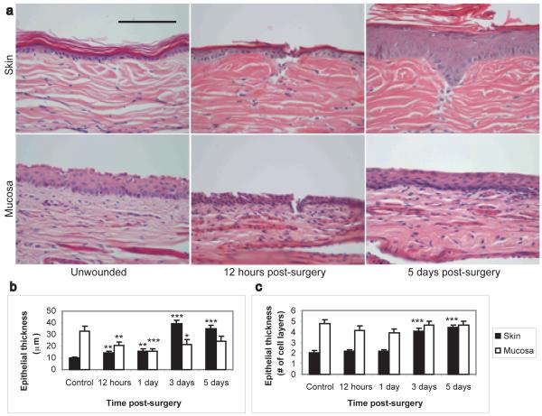
Figure 1. Differential epithelial response to wounding in skin and mucosa.
Representative histology photomicrographs of control and injured skin and vaginal mucosa (A) demonstrate that following injury, the cutaneous epithelium undergoes significant hypertrophy while the mucosal epithelium does not. Measurement of epithelial thickness (B and C) indicates that mucosal wounds heal via restitution, resulting in a transient thinning of the epithelium without a reduction in the number of cell layers. In contrast, cutaneous wounds healed by keratinocyte proliferation, resulting in a significant increase in epidermal thickness following re-epithelialization. Magnification: 40X. Scale bar = 100μm. N=8 wounds per time point. * = p<0.05, ** = p<0.01, *** = p<0.001.

