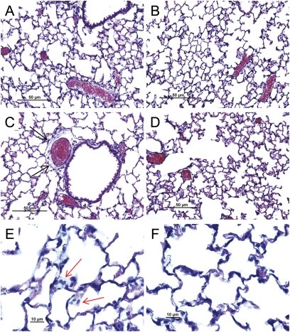
Figure 2.
Histological assessment of lung tissue from (A) control SM/J mice, (B) control 129X1/SvJ mice, (C, E) acrolein-exposed SM/J mice, or (D, F) acrolein-exposed 129X1/SvJ mice. Consistent with acute lung injury, (C) perivascular enlargement (black arrow) and (E) leukocyte infiltration (red arrow) were more evident in the (C, E) sensitive SM/J strain than in the (D, F) resistant 129X1/SvJ strain. Mice were exposed to filtered air (control) or to acrolein (10 ppm, 17 h) and killed. Lung tissue was obtained, fixed in formaldehyde, and 5-μm sections prepared with hematoxylin and eosin stain. Bars indicate magnification.

