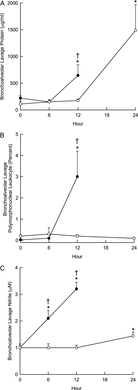
Figure 3.
Characterization of acute lung injury by bronchoalveolar lavage. Mice were exposed to 10 ppm acrolein for 0 (filtered air control), 6, or 12 hours, killed, and bronchoalveolar lavage performed with (Ca2+, Mg2+ free) phosphate buffered saline. (A) Bronchoalveolar lavage protein increased sooner in the sensitive (SM/J) than in the resistant (129X1/SvJ) mouse strain. Lavage fluid was centrifuged and total protein in cell-free supernatants was measured using a bicinchoninic acid assay. (B) Bronchoalveolar lavage polymorphonuclear leukocytes increased in the sensitive (SM/J) but not in the resistant (129X1/SvJ) mouse strain. After centrifugation, cell pellet was suspended and an aliquot (200 μl) were cytocentrifuged and the cells were stained with Hemacolor for differential cell analysis according to standard cytological procedures. (C) Bronchoalveolar lavage nitrite concentration increased sooner in the sensitive (SM/J) than in the resistant (129X1/SvJ) mouse strain. Supernatant was analyzed using a fluorometric method in which nitrite reacted with 2,3-diaminonaphthalene. *Significantly different from strain-matched control as determined by analysis of variance with an all pairwise multiple comparison procedure (Holm-Sidak method). †Significantly different between the sensitive SM/J and resistant 129X1/SvJ mouse strain as determined by analysis of variance with an all pairwise multiple comparison procedure (Holm-Sidak method).

