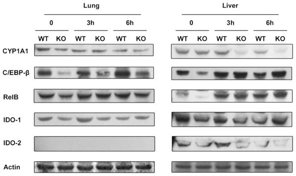
Fig. 2.
Protein levels of CYP1A1, CEBPβ, RelB, IDO-1, and IDO-2 after LPS treatment. Protein levels of selected AhR and LPS target genes in the lung and liver tissue of wild-type (WT) and AhR−/− (KO) mice were detected using Western blot analysis. Whole tissue protein from the lung and liver tissue of WT and KO mice was used for analysis. A Western blot of representative samples from each group is shown. The expression level of β-actin was used as a loading control.

