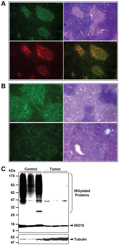
Figure 2. Expression of ISG15/ISGylation in K-rasLA2 thymic tumors.
A&B, Representative immunofluorescence images showing ISG15 expression in normal and cancerous mouse thymuses. A. 100×, ISG15 expression is predominantly localized to the medulla of a normal thymus. Top left, ISG15 staining; top right, H&E staining of the adjacent frozen section; bottom left, staining of a medullary marker (G8.8); bottom right, merged image of ISG15 and G8.8 staining. B. 200×, ISG15 expression in a normal (top) and cancerous thymus (bottom). Images on the right are H&E staining of adjacent tissue sections. C. Western blotting analysis of ISG15 and protein ISGylation levels in normal (lanes 1–3) and cancerous (lanes 4–6) thymic tissues. The membrane was blotted with anti-tubulin α antibody first before it was striped and reprobed with anti-ISG15 antibody.

