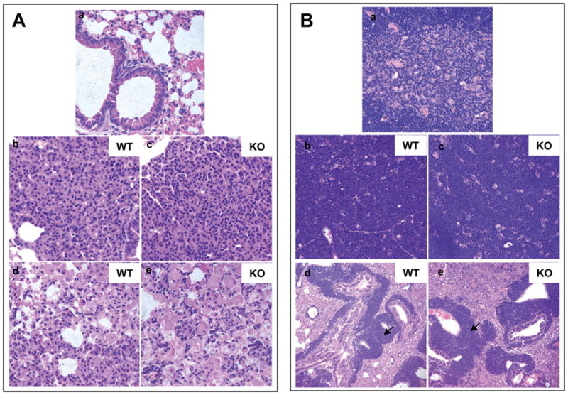
Figure 4. Ube1l+/+/K-rasLA2 and Ube1l−/−/K-rasLA2 mice exhibited similar tumor pathology.

A. H&E staining images of lung tissues. a. A normal lung from a wild type mouse. b&c. Lung alveolar adenomas from 12-week old mice. d&e. Lung lesions in moribund mice showing irregular cell shapes. B. H&E staining images of normal thymuses and thymic lymphomas. a. A normal thymus showing the medulla structure. b&c. A thymus with lymphoma showing a sheet of lymphocytes with scattered star-like cells. d&e. Infiltration of lymphoma cells (indicated by arrows) in the lung. b&d are representative images for Ube1l+/+/K-rasLA2 mice. c&e are representative images for Ube1l−/−/K-rasLA2 mice. All images are at 400× magnification except figure B-(d) and figure B-(e), which are at 100× magnification.
