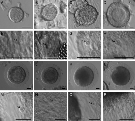
Figure 1.
Morphology of intraovarian cat oocytes (A–D and I–K) and respective GVs delimited by a nuclear envelope (E–H and M–O) at different stages of follicular development. (A and E) Primordial. (B and F) Primary. (C and G) Secondary. (D and H) Pre-antral. (I and M) Early antral. (J and N) Small antral. (K and O) Large antral. (L and P) After artificial chromatin compaction. Black arrows indicate the presence of the nucleolar-like body. Bar = 20 µm.

