Figure 3.
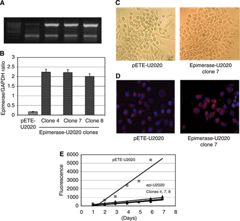
Ectopic expression of GLCE in U2020 small-cell lung cancer cells. (A) Representative multiplex RT–PCR gel showing GLCE expression in U2020 cells stably transfected with the pETE or epi-pETE plasmids (clones 4, 7 and 8). (B) GLCE expression levels normalised to that of GAPDH. The graph shows the mean expression levels from triplicate experiments (±s.d.) (OriginPro 8.1). (C) Morphology of stably transfected pETE-U2020 cells and epi-U2020 cells (clone 7); magnification × 100. (D) Immunocytochemical analysis of GLCE expression in pETE-U2020 and epimerase-U2020 cells using a custom anti-epimerase polyclonal antibody. (E) Proliferation rates of the stable epi-U2020 and control pETE-U2020 clones (CyQUANT NF Cell proliferation assay).

