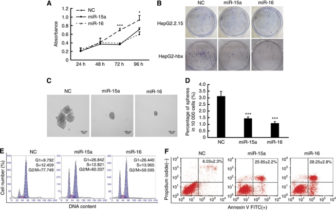
Figure 5.
Ectopically expressed miR-15a/16 repressed the proliferation, clonogenicity, and anchorage-independent growth of HBx-transfected HepG2 cells in vitro by blocking cell-cycle progression and inducing apoptosis. (A) CCK-8 analysis showed that the expression of both miR-15a and miR-16 significantly suppressed the growth of HepG2-hbx cells at 72 and 96 h post-transfection. The data are presented as mean absorbance±s.e. (*P<0.05, ***P<0.001, n=3; Student's t-test). (B) HepG2-hbx and HepG2.2.15 cells transfected with miR-15a/16 or NC were grown at an extremely low density (e.g., 500–1000 cells in 10 ml of medium) for 2–3 weeks, and the clones were fixed with methanol and stained with 0.1% crystal violet in 20% methanol. Representative plates are presented. (C and D) HepG2-hbx cells (10 000) transfected with miR-15a/16 mimic or NC were placed in a 10-ml sphere formation assay-conditioned medium to grow anchorage independently for 10 days. The data are presented as the mean numbers of spheres ±s.e. (***P<0.001; n=3, Student's t-test). (E) HepG2-hbx cells were treated with nocodazole (100 ng ml−1) 24 h post-transfection, and the DNA ploidy was analysed 20 h later. 2N, cells containing diploid DNA; 4N, cells containing tetraploid DNA. (F) PI-Annexin V analysis was performed to detect the early apoptosis of HepG2-hbx cells transfected with miR-15a/16 or NC (48 h). The data are presented as mean±s.e. (n=3).

