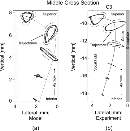
Figure 8.
(a) Five simulated 3D trajectories (dotted lines) along middle vertical cross section produced by 3DM. (b) Six experimental 3D trajectories (dotted lines) along middle vertical cross section extracted from an excised human hemilarynx (Ref. 26). The vertical contours in both images correspond to the rest positions of the mass elements as well as the vocal fold tissue, respectively. For all five mass elements (from inferior up to superior) in (a) the movement ranges can be roughly described as 0.5, 1, 2, 2, and 1.5 mm, respectively. By comparison, the movement ranges of the upper five suture points (from inferior up to the vocal fold edge) in (b) are almost 0.3, 1, 2, 2, and 1.7 mm, respectively. Hence, the trajectories show similar behavior.

