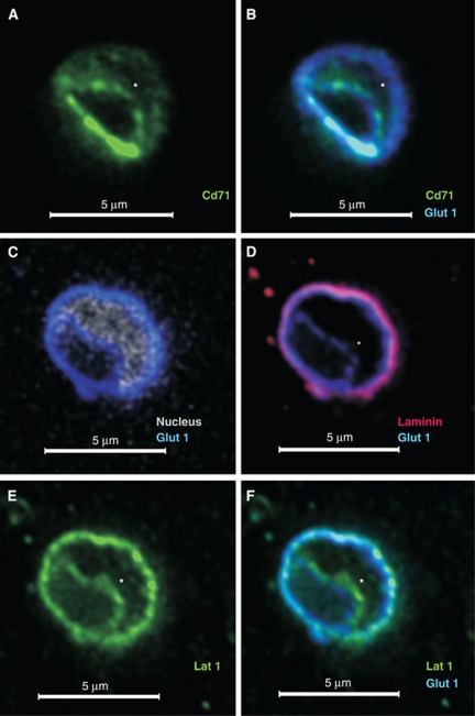
Figure 1.
Localization of specific markers at the luminal and/or abluminal membrane of the brain vasculature. Freshly PFA-fixed mouse brain tissue sections stained as indicated were imaged using confocal microscopy. Representative images of ∼5-μm-diameter capillaries stained with: (A, B) anti-Cd71 (green; marker for the luminal membrane), anti-Glut1 (blue; marker for both membranes) or (C–F) with antibodies against Lat1 (green; known to be expressed on both membranes), Glut1 (blue), laminin (red; marker for extracellular matrix separating the abluminal membrane from brain cells), and the nuclear dye PO-PRO (white; separates the luminal from the abluminal membrane). The nucleus is indicated by ‘*' if PO-PRO nuclear staining is not shown. PFA, paraformaldehyde.

