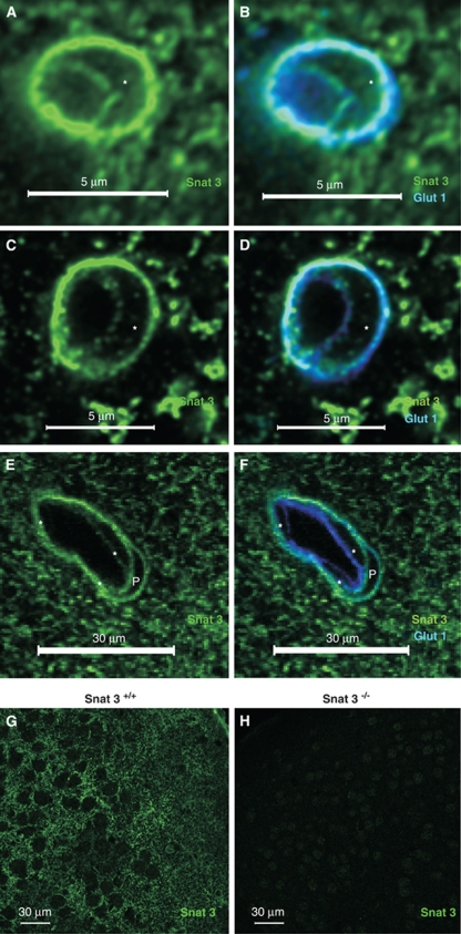
Figure 3.
Snat3 is localized on both BBB endothelial membranes. (A, B) Representative confocal image of a capillary from fresh fixed mouse brain sections stained with anti-Snat3 (green) and anti-Glut1 (blue), and the nucleus is indicated by ‘*', showing colocalization of Snat3 with Glut1 on both BBB membranes in adult (5-week-old) wild-type mice. (C–F) As Snat3−/− mice generally die before weaning, the analysis was carried out using tissue obtained from postnatal day 10 mice. Representative confocal images of tissue sections from 10-day-old heterozygous (Snat3+/−) mice showing (panels C and D) a capillary and (panels E and F) a large microvessel stained with antibodies against Snat3 (green), Glut1 (blue), or nuclear dye, PO-PRO (white), as indicated, verifying the expression of Snat3 in cortical microvessels of different sizes. Endothelial nuclei were indicated by ‘*' and the pericyte nucleus was designated by ‘P'. (G, H) Representative overview confocal images stained with Snat3 antibody (green) verifies expression of Snat3 in brain parenchyma from 10 day wild-type (Snat3+/+; panel G) but not knockout (Snat3−/−; panel H) mice. BBB, blood–brain barrier.

