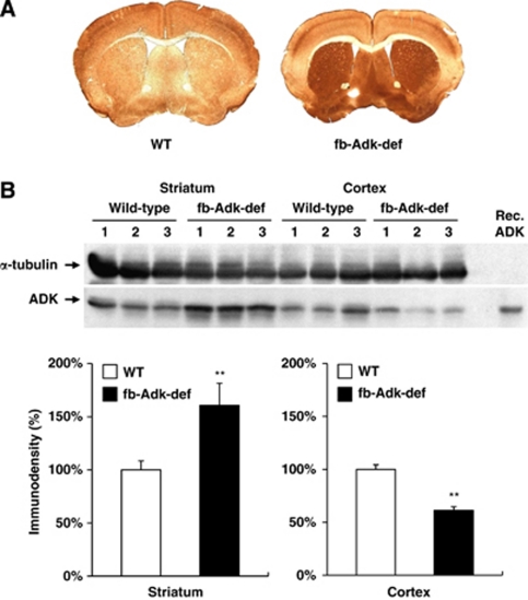
Figure 1.
Differential expression pattern of striatal and cortical adenosine kinase (ADK) in fb-Adk-def mice. Regional expression of ADK was evaluated in the brain of naive wild-type (WT) and fb-Adk-def mice. (A) Representative immunohistochemical staining with ADK primary antibody in WT (left) and fb-Adk-def (right) mice. (B) (Top) Representative Western blot of ADK from the striatum or cortex of adult WT and fb-Adk-def mutant mice. (Bottom) Quantitative analysis of striatal (left panel) and cortical (right panel) ADK levels based on two replicates of Western blots performed with samples from n=5 animals from each genotype. ADK levels were first normalized for loading using a α-tubulin standard. ADK levels are shown as relative to striatal or cortical ADK levels in WT mice (set as 100%). Data are displayed as mean±s.e.m. **P<0.01 paired comparisons t-test.

