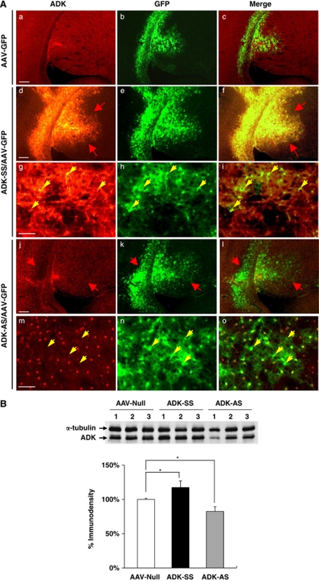
Figure 5.
Modulation of adenosine kinase (ADK) expression with an adeno-associated virus (AAV)-based vector system. (A) Immunohistofluorescence of ADK (red) and green fluorescent protein (GFP) (green) in wild-type (WT) mice injected with AAV-GFP (a–c), or coinjection of AAV-GFP with either ADK-SS (d–i), or ADK-AS (j–o). (a–c) Representative immunohistofluorescence showing basal ADK levels and AAV-virus expression pattern in AAV-GFP-injected WT mice. (d–f) ADK-SS/AAV-GFP coinjection causes a robust increase in ADK immunoreactivity (red arrows). (g–i) Higher magnification images of the ADK-SS/AAV-GFP coinjection site shows that ADK colocalizes with AAV-GFP-infected cells (yellow arrows). (j–l) ADK-AS/AAV-GFP coinjection in WT mice causes a decrease in ADK immunoreactivity (red arrows). (m–o) Higher magnification images of the ADK-AS/AAV-GFP coinjection site show that cells lacking ADK colocalize with AAV-GFP-infected cells (yellow arrows). (B) (Top) Representative Western blot of ADK from adult WT mice injected with AAV-null, ADK-SS, or ADK-AS virus. (Bottom) Quantitative analysis of ADK levels based on two separate Western blots performed with samples from n=4 animals for each injection type. Protein loading was normalized to the α-tubulin standard before intergroup comparison. Values are displayed as relative to ADK protein levels in AAV-null (set as 100%) brain. Data represent the mean±s.e.m., n=4. *P<0.05 versus AAV-null group.

