Figure 1.
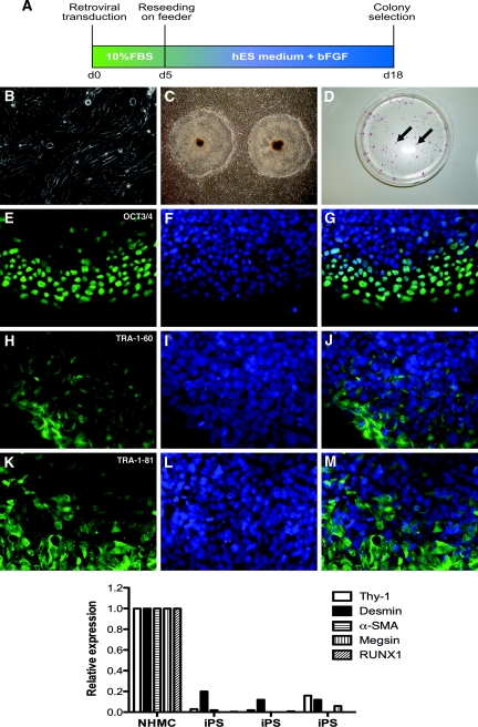
Kidney mesangial cell-derived iPS cells express stem cell markers. (A) The timeline of induction of iPS cells from human mesangial cells following retroviral transduction. (B) Representative images of normal cultured human mesangial cells reprogrammed to generate iPS colonies (C). (D) Alkaline phosphatase-positive iPS colonies (arrows). Immunofluorescence staining of mesangial cell-derived iPS colonies shows localization of OCT3/4 protein (E; green), with corresponding DAPI-stained nuclei (F; blue) and a merged image (G). TRA-1-60 (H–J) and TRA-1-81 (K–M) proteins are also expressed. Original magnifications (B, E–M ×400, C ×10). The graph in the lower panel shows qPCR of mesangial cell markers in NHMCs, with a downregulated expression in iPS cells from 3 separate colonies at passage 4. 120 × 189mm (600 × 600 DPI).

