Figure 7.
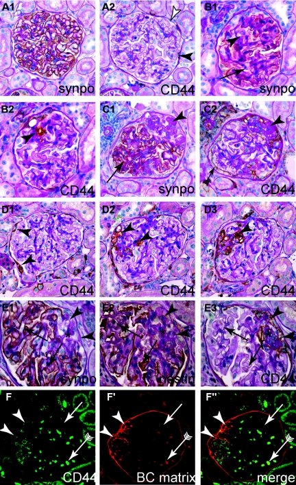
Activated parietal cells are present within sclerotic lesions of the MWF rat. (A through C) Representative serial sections of aged female MWF rats, consecutively stained for synaptopodin as podocyte marker or CD44 as marker for activated PECs. (A1 and A2) Normal glomerulus. Although most PECs are CD44-negative (white arrowhead), focal CD44 expression can be observed in PECs (black arrowhead). (B1 and B2) A small sclerotic lesion, where synaptopodin staining is reduced and replaced by CD44-positive cells (arrowhead). (C1 and C2) Advanced sclerotic lesion, where podocytes are absent (arrowhead) and replaced by matrix or CD44-positive cells. Note that CD44-positive cells on BC show a more activated phenotype (more cuboid, arrow with tale). (D1 through D3) Serial sections of a glomerulus affected by a segmental sclerotic lesion (arrowheads D2 and D3). Note that on section D1 the lesion cannot be seen. Focally activated CD44-positive PECs are the only sign of a sclerotic lesion in another plane of sectioning (arrowheads, D1). (E1 through E3) To exclude a potential loss of the podocyte marker synaptopodin, serial sections were also stained for the podocyte marker nestin. Nestin always colocalized with synaptopodin (arrows). Nestin was not expressed in sclerotic lesions (asterisk), which was populated by CD44-positive cells [arrowheads; (A through E) immunohistologic stainings on paraffin sections counterstained with PAS]. (F through F″) BC-type matrix was deposited in sclerotic lesions (arrowheads, CD44-positive) but not along the intact glomerular tuft (arrow). Erythrocytes show autofluorescence in all channels (arrow with tails; immunofluorescent double staining on paraffin sections).

