Figure 9.
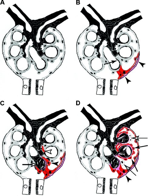
Schematic summary of the role of parietal cells in glomerulosclerosis. (A) A normal glomerulus consists of the endocapillary compartment (capillaries and mesangium in black) and epithelial cells: podocytes lining the capillary tuft (gray cells) and PECs lining the inside of BC (white cells). (B) Focal activation of PECs (red) and formation of an adhesion to the capillary are the first events in the formation of a sclerotic lesion. In some cases, focally activated PECs also produced more matrix on BC (pink matrix). Podocytes in the immediate vicinity of an adhesion are often effaced. (C) Activated PECs invade the affected segment of the glomerular tuft (black arrowhead) and deposit BC-type matrix. The invading PECs remain strictly within the extracapillary compartment. Occasionally, they appear disconnected from the adhesion due to tangential sections (white arrowhead). Within the endocapillary compartment, mesangial sclerosis develops within the affected segment (white arrow). Sclerosis of the glomerular tuft progresses from the adhesion. (D) Advanced sclerotic lesion. The former capillary loops are covered by parietal cells (black arrows) and BC-type matrix (pink). In advanced lesions, some invading parietal cells no longer express markers of activation (white arrows). At the site of adhesion, a continuous bridge of BC-type matrix has formed between Bowman's capsule and the glomerular tuft (black arrowhead).

