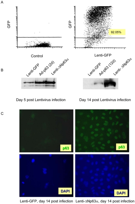
Figure 1. Lentivirus infection drives long term and stable gene expression in primary keratinocytes.
A, Primary mouse keratinocytes were infected with lentivirus encoding GFP, and infection efficiency was assessed by FACS analysis at day 5 post-infection. Y-axis indicates the intensity of GFP, X-axis represents forward angle light scatter. B, Primary keratinocytes were infected with lentivirus encoding GFP or ΔNp63α and whole cell protein was collected at day 5 and 14 and analyzed for p63 expression by western blot. C, Primary keratinocyte cultures expressing lenti-GFP or lenti-ΔNp63α were fixed at 14 days post-lentiviral transduction and incubated with anti-p63 antibody, followed by a secondary antibody conjugated with Alexa-488. The cells were then stained with DAPI to visualize the cell nuclei.

