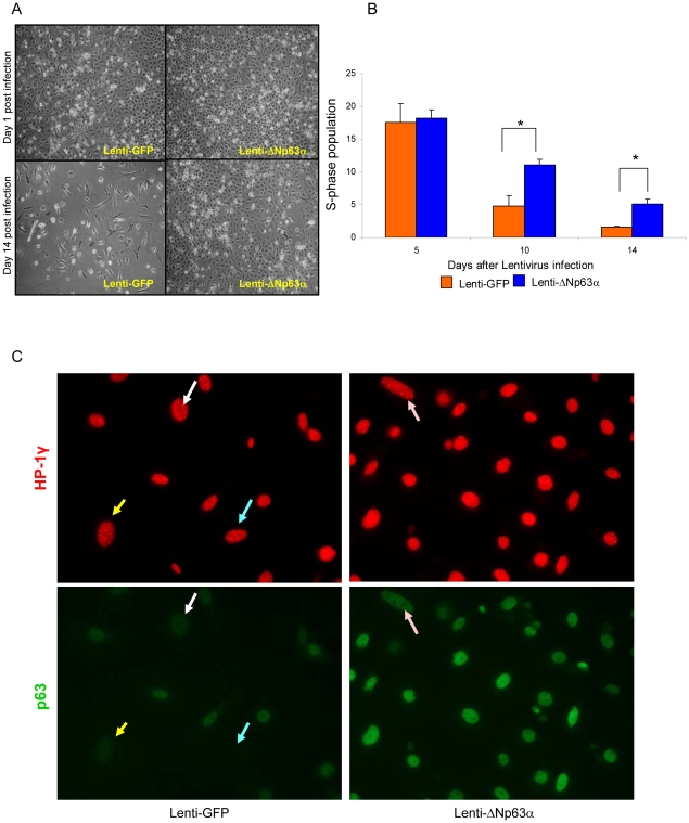
Figure 2. Dysregulated expression of ΔNp63α promotes survival and blocks replicative senescence of primary keratinocytes.
A, Phase morphology of lenti-GFP- and lenti-ΔNp63α expressing keratinocytes cultured for 1 and 14 days post-infection. Results shown are representative of three independent experiments. B, Quantification of BrdU positive cells in lenti-GFP or lenti-ΔNp63α cultures at timepoints noted following lentiviral gene transduction. Data shown represent the means ± S.E. of three independent experiments. * indicates a statistically significant difference between lenti-GFP and lenti-ΔNp63α infected cells at p<0.05. C, Primary keratinocytes expressing lenti-GFP (left panel) or lenti-ΔNp63α (right panel) were fixed at 14 days post-lentiviral transduction. The cell senescence status was assessed by the presence of HP-1γ nuclear foci (red). The cells were double stained with p63 antibodies (green). Matched arrows indicate the same cell stained with both different antibodies. The image presented is representative of three independent experiments.

