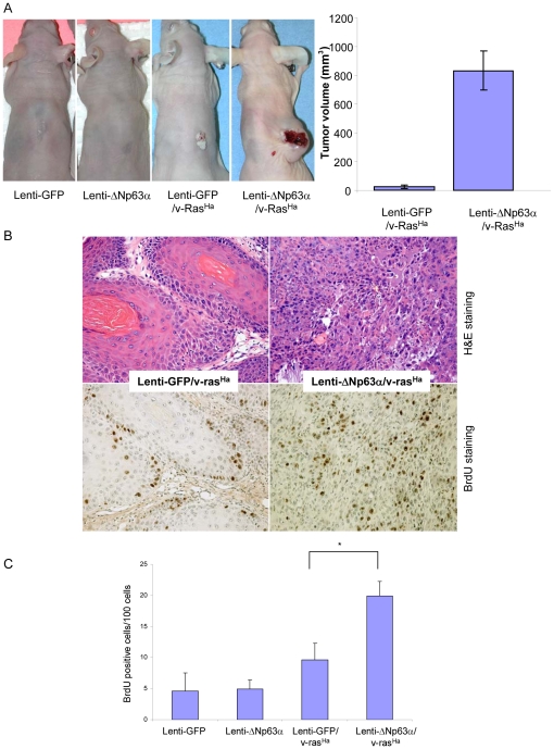
Figure 4. Elevated expression of ΔNp63α enhances malignant conversion in v-rasHa-expressing keratinocytes.
A, In vivo phenotype of ΔNp63α-overexpressing keratinocytes in the presence and absence of oncogenic ras. Primary mouse keratinocytes were infected with lentivirus encoding lenti-GFP or lenti-ΔNp63α or sequentially with retrovirus encoding v-rasHa followed by lenti-GFP or lenti-ΔNp63α. The final tumor phenotype was assessed at 5 weeks following grafting. Final tumor volumes at 4 weeks are presented as the mean tumor volume ± S.E. B, Upper panel, representative H&E sections of tumor tissues obtained from grafting sites. Lower panel, BrdU incorporation in grafted tissue samples. C, Quantification of BrdU incorporation levels. Data are presented as the mean % of BrdU positive cells ± S.E. * indicates a statistically significant difference between lenti-GFP/v-rasHa and lenti-ΔNp63α/v-rasHa groups at p<0.05.

