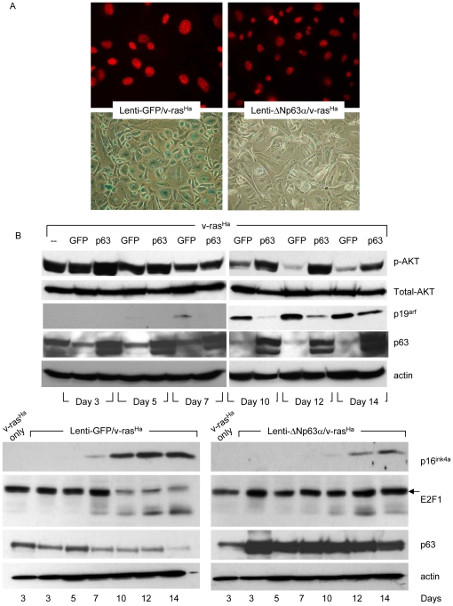
Figure 5. Long term ΔNp63α overexpression blocks oncogene-induced senescence.
A, Primary keratinocytes were infected sequentially with retrovirus encoding oncogenic v-rasHa, followed by lentivirus encoding GFP or ΔNp63α. Oncogene-induced senescence was assessed 14 days post-lentivirus infection by immunofluorescent analysis of nuclear foci of HP-1γ (upper panel), or enzymatic activity of SA-β-gal (lower panel). The images shown are representative of three independent experiments. B, Whole cell protein was collected from keratinocytes infected sequentially with retrovirus encoding oncogenic v-rasHa followed by lentivirus encoding GFP or ΔNp63α at timepoints indicated following lentiviral gene transduction. The levels of phosphorylated AKT, total AKT, p19arf and p63 were detected by western blot (upper panels). p16ink4a and E2F1 levels with corresponding p63 expression are presented in the lower panels. Equal protein loading was confirmed by immunoblotting for β-actin.

