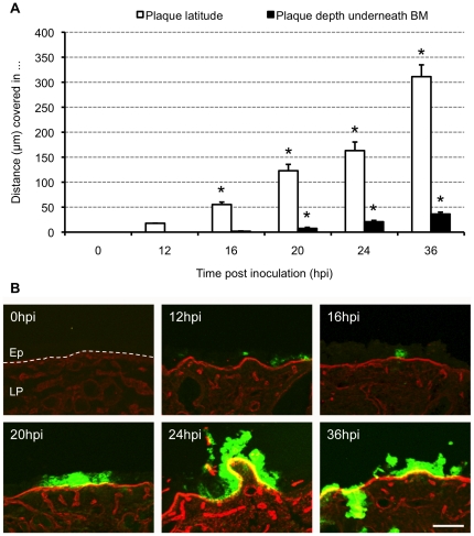
Figure 4. Evolution of HSV1 mucosal spread.
(A) Kinetic evolution of HSV1 plaque formation. Explants were inoculated with 1 ml virus suspension containing 107 TCID50/ml HSV1 VR-733 and sampled at 0, 12, 16, 20, 24 and 36 h post inoculation (pi). Serial 20 µm cryosections were made and plaque latitude (white bars) and plaque depth underneath the basement membrane (BM), distance covered by HSV1 in the lamina propria, (black bars) were measured using ImageJ. Data are represented as means of 10 plaques of triplicate independent experiments+SD (error bars). *, Significant differences at the 0.05 level. (B) Representative confocal photomicrographs of the evolution of HSV1 VR-733 spread in human nasal respiratory explants at 0, 12, 16, 20, 24 and 36 h pi. Collagen IV is visualised by red fluorescence. Green fluorescence visualises HSV1 antigens. Bar, 100 µm. Abbreviations: Ep, epithelium; LP, lamina propria. The BM is marked with a dashed line.

