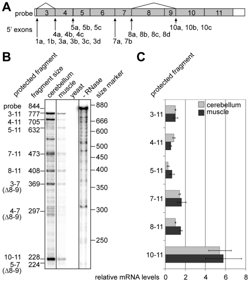
Figure 2. Initiation of transcription from alternative sites within the TCF4 gene.
(A) Schematic representation of the ribonuclease protection assay probe complementary to TCF4 exons 3–11. Location of the TCF4 5′ exons relative to the probe is shown with arrows and the sites of alternative splicing with lines. (B) Autoradiograph of the probe fragments protected by human cerebellum or muscle RNA and fragments obtained from control reactions with yeast RNA or without RNase treatment. The expected sizes of the protected fragments in bps and the exons they span are shown at the left and the location of the size markers at the right. (C) Densitometric quantification of the protected fragments in B from two assays. The values are given in relation to the levels of the fragment spanning exons 3–11 for both tissues. Error bars indicate standard deviations.

