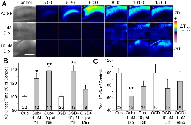
Figure 2. Dibucaine pretreatment delays terminal depolarization in human neocortical slices.
A, Changes in LT (ΔLT) are utilized to track terminal depolarization in real time. The first image in each row shows bright field image of the slice. A control untreated slice incubated in standard ACSF (top row) undergoes terminal depolarization within 5 minutes of superfusion with 100 µM ouabain. Pseudocolored elevated ΔLT indicates cell swelling. In slices pretreated with either 1 or 10 µM dibucaine for 60 min (middle and bottom rows, respectively), ouabain-induced terminal depolarization is delayed by 2.1 and 4.8 min respectively and has less impact in terms of cell swelling. All three slices were from the same patient (#7; see Patient Table S1). Scale bar, 1 mm. B, Pretreatment for 1 hour with dibucaine significantly increases the latency to terminal depolarization onset induced by 100 µM ouabain or OGD when compared to untreated control slices from the same patient. C, Peak cell swelling during terminal depolarization in the same slices pretreated with dibucaine. The neuroprotective anti-inflammatory drug minocycline had no effect on terminal depolarization. Numbers of slices in each condition are indicated within each bar. Values are shown as percent of control (untreated slices from the same patient). Asterisks indicate significant differences from control (*p<0.05, **p<0.001, two-way ANOVA).

