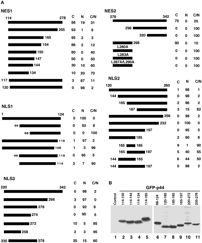
Figure 3. Mapping of nuclear transport signals in the p44/WDR77 molecule.
(A) Diagrams of the p44/WDR77 truncations expressed as GFP-fusion proteins in Cos 7 cells. The percentages of cells with GFP-p44/WDR77 truncations in cytoplasm (C), nucleus (N), or cytoplasm plus nucleus (C/N) are shown on the right. (B) Western blot analysis of whole-cell extracts derived from Cos 7 cells transfected with pcDNA-f:GFP-p44/WDR77 truncations (lanes 2–11) with anti-FLAG antibody. Lane 1 is the whole-cell lysate from nontransfected Cos 7 cells.

