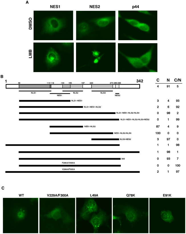
Figure 6. The relationship between identified nuclear export and import signals.
(A) LMB does not inhibit translocation of p44/WDR77 to the cytoplasm. Cos 7 cells were transfected with pcDNA-GFP-NES1 or -NES2 (left two panels). The transfected cells were grown in the presence of dimethylsulfoxide (DMSO) or 20 mM of LMB for 6 hr. Subcellular localization of GFP-NES1 and GSP-NES2 was observed under a confocal microscope. Cos 7 cells were grown in the presence of DMSO or 20 µM of LMB for 6 hr and immunostained with the anti-p44 antibody (right panel). (B) Cos 7 cells were transfected with constructs expressing GFP-fusion proteins of various p44/WDR77 truncations or point mutations as indicated. The transfected cells were stained with TO-PRO 3 and observed under a confocal microscope. The percentages of cells with GFP signals in cytoplasm (C), nucleus (N), or cytoplasm plus nucleus (C/N) are shown on the right. (C) Point mutations on p44/WDR77 abolished its nuclear localization in Cos 7 cells. Cells were transfected with constructs expressing the wild-type or mutant (V299A/F300A, L49A, Q78K, or E91K) GFP-p44/WDR77, stained with TO-PRO 3, and observed under a confocal microscope.

