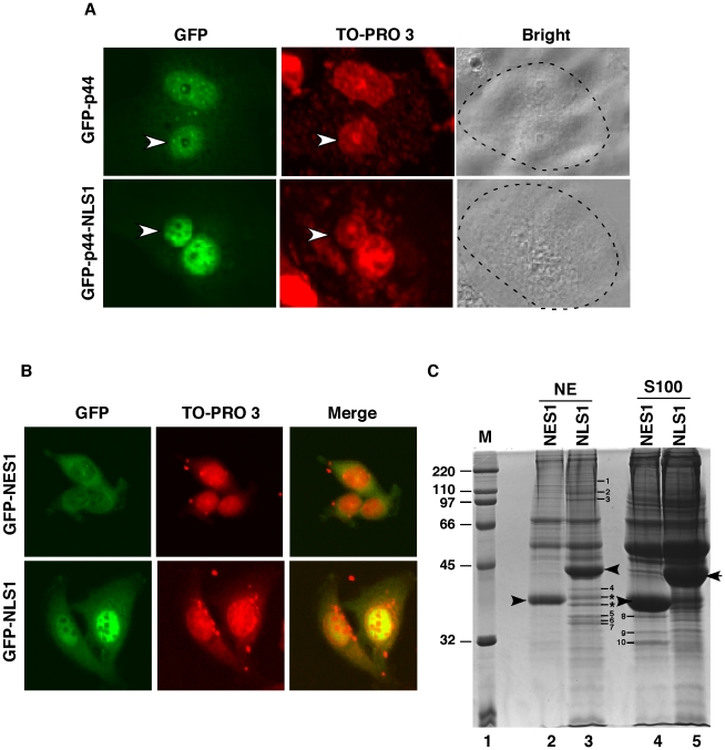
Figure 8. Cos 7 cells restore p44/WDR77 nuclear localization in LNCaP cells.
(A) LNCAP cells were transfected with pcDNA-GFP-p44/WDR77 or pcDNA-GFP-NLS1. After expression of the transfected DNAs, the cells were fused to Cos 7 cells to form heterokaryons. After incubation for 6 hr, the cells were fixed and counterstained with TO-PRO 3, which differentiates the human and monkey nuclei within the heterokaryon (arrows identify the human nuclei). The panel marked Bright shows the phase-contrast image of the heterokaryons; the broken line highlights the cytoplasmic edge. (B) Cytoplasmic localization of f:GFP-NES1 and nuclear localization of f:GFP-NLS1 in PC3 cells. PC3 cells stably expressing f:GFP-NES1 (top panels) or f:GFP-NLS1 (bottom panels) were stained with TO-PRO 3 and observed under a confocal microscope. (C) Identification of polypeptides that were associated with NES1 or NLS1. The bands corresponding to f:GFP-NES1 and f:GFP-NLS1 are indicated by arrows. Polypeptides specifically associated with f:GFP-NLS1 or f:GFP-NES are indicated by numbers and stars. The standard protein markers (Bio-Rad) are shown in lane 1.

