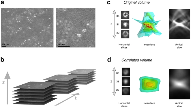
Figure 2. Cell appearance in 3D collagen gels.
(a) Cell and collagen gel aspects in an original phase-contrast Z-slice with a zoom showing the collagen fibers. (b) Schematic presentation of the 3D time-lapse sequence acquisition. Detailed cell aspect in (c) the original phase-contrast volume (after contrast enhancement) and (d) the correlated volume, illustrated by several Z-slices, a XZ cut plane (vertical slice) and an intensity isosurface 3D view.

