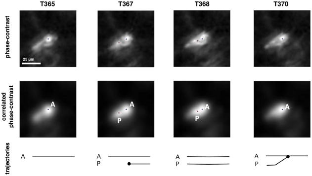
Figure 6. Tracking of an elongated cell.
An elongated cell (blue dot) spawns a second parasite tracker (red dot) on a body extension (on the cell tail before its retraction when migrating). The secondary tracker is discarded as it converges onto the cell's original tracker within a couple of frames. The images show Z-slices centered on the cell in the phase-contrast (1st row) and correlated (2nd row) volumes. The timeline below the images is similar to that in figure 4.

