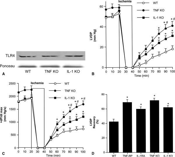
Fig 3.
Role of tumor necrosis factor (TNF)-α and interleukin (IL)-1β in postischemic cardiac dysfunction. (A) Immunoblotting demonstrates that TNF-α knockout (TNF KO), IL-1β knockout (IL-1 KO) and wild-type (WT) hearts have comparable Toll-like receptor 4 (TLR4) levels. (B, C) Hearts isolated from TNF KO (black circles), IL-1 KO (black squares) and WT (white circles, a combined group of TNF-α WT and IL-1β WT) underwent 20 minutes of global ischemia followed by 60 minutes reperfusion. TNF KO and IL-1 KO hearts had higher left ventricular developed pressure (LVDP) and +dP/dt max after I/R than hearts from wild-type controls. Data are expressed as mean ± SEM. n = 6 in each group; *p < 0.05 vs WT; #p < 0.05 vs IL-1 KO. (D) Wild-type hearts underwent 20 minutes of global ischemia followed by 60 minutes reperfusion, and were treated with TNF-binding protein (TNF-BP, 1.0 μg/mL) or IL-1 receptor antagonist (IL-1 RA, 1.0 μg/mL) during reperfusion. The percentage recovery of LVDP in hearts treated with TNF-BP and IL-1 RA was greater than in untreated hearts and was similar to knockouts. Data are expressed as mean ± standard error of the mean; n = 6 in each group; *p < 0.05 vs WT.

