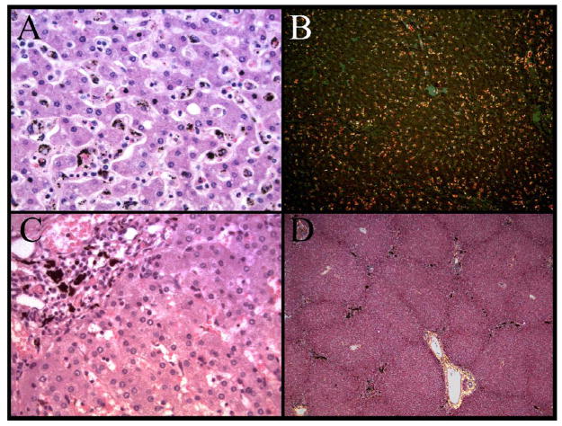
Figure 2.
The distribution of pigment within the liver of the autopsy cohort showed two distinct patterns. A diffuse, speckled pattern predominantly in the lobular parenchyma was present and associated with increased histiocytes within sinusoids (A). At low power, polarized microscopy reveals the extent of the histiocyte hyperplasia (B). Within portal triads, large globules of pigment were present in many cases (C). At low power, the lobular architecture and the increased pigment (present as black areas) in the portal triads is evident (D).

