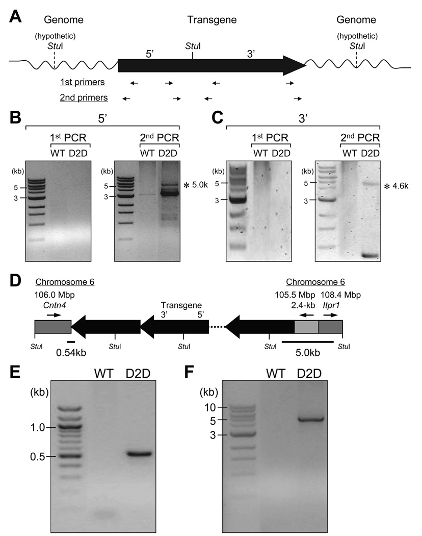
Figure 2.
Identification of the genomic DNA sequences flanking the transgene in D2D mice. Strategy of the inverse PCR (IPCR). The D2/LacZ transgene is presented with 5’ to 3’ orientation. The single StuI restriction site on the transgene is indicated by a solid line, with the assumed ones on the flanking genomic DNA represented by dash lines. The restrictive DNA fragments were ligated to form circles. The position and orientation of the nested PCR primers are indicated by arrows underneath the transgene, for detection of the genomic sequences flanking either the 5’-end or the 3’-end of the transgene (A). Agarose gel electrophoresis of the products from the nested PCR. Asterisks indicate the PCR products containing mouse genomic DNA, flanking the 5’-end (B) or the 3’-end (C) of the transgene, as revealed by sequencing and BLAST with assembled mouse genome. Schematic drawing of the transgene tandem repeats with flanking genomic sequences on mouse chromosome 6, as suggested by IPCR. The gray boxes represent the genomic DNA, with arrows above indicating their 5’ to 3’ orientation on the chromosome 6. Their positions on the chromosome and the involved loci are also noted on top. The StuI sites on the transgene repeats and the flanking DNA are labeled (D). To independently determine the junction sequences, the primer pair M106M-f6 and LacZ-3F was used for the 3’-end of the transgene, yielding a 0.54-kb PCR product (E) and the G78P-1R and ItprR pair was used for the 5’-end, yielding a 5.0-kb PCR product (F).

