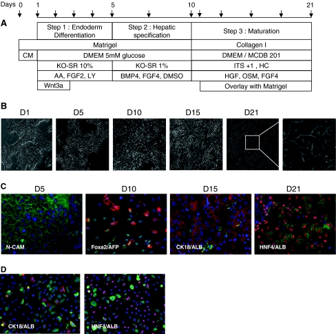
Fig. 1.
Differentiation of hES cells. A) Protocol. Arrows indicate medium renewal. B) Photomicrographs of morphological changes during differentiation. C) and D) immunofluorescence analysis of N-CAM, FOXA2, AFP, HNF4, CK8-18 and Albumin in ES-Hep and PCHH. Only overlays (FOXA2-green/AFP-red; ALB-green/CK8-18 or HNF4-red) are shown here. See Supplementary Fig. 2 for further detail

