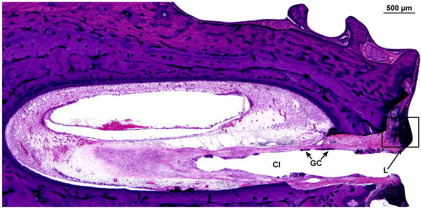
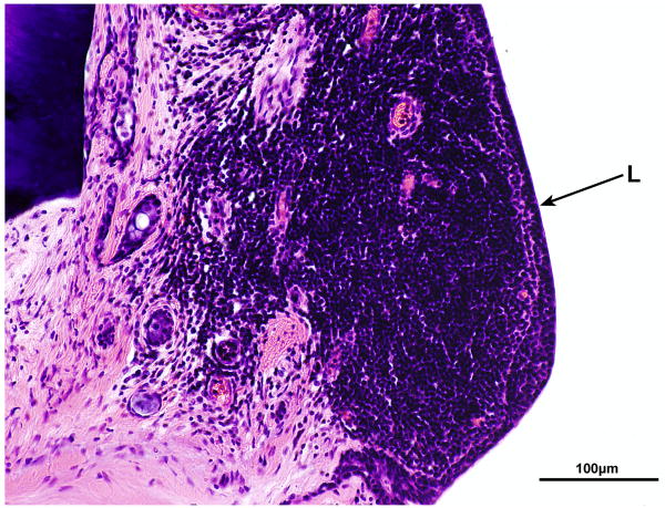
FIG. 11.
Right cochlea. A, Section of the basal turn near the cochleostomy site. There was an intense fibrous reaction and foreign body giant cells (GC) and lymphocytes (L) around the cochlear implant (CI) as it entered the scala tympani. B, High power of boxed area shown in Figure 11A. An intense infiltrate of lymphocytes (L) was adjacent to the implant track.


