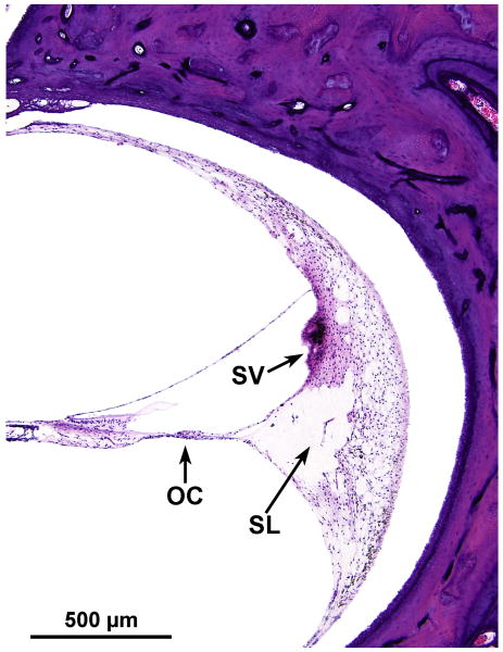
FIG. 3.
Higher power photomicrograph of the basal turn of the organ of Corti. This demonstrated severe degeneration of the organ of Corti (OC) and stria vascularis (SV) and severe atrophy of the spiral ligament (SL), especially in the area of its attachment to the basilar membrane, perhaps resulting in artifactual separation of the spiral ligament from the lateral cochlear bony wall. Granular inclusions were common within the stria.

