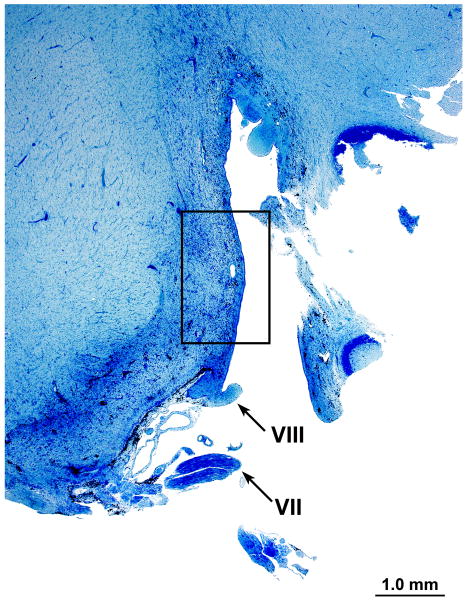
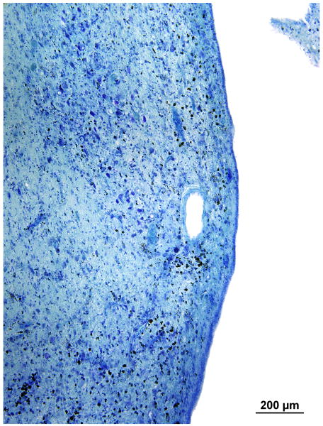
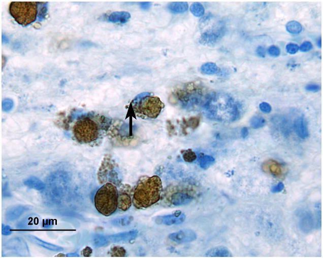
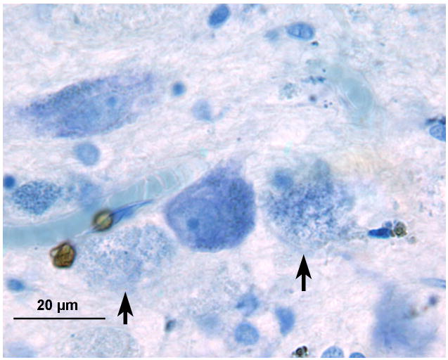
FIG. 5.
Left cochlear nucleus. Azure stain for nucleic acid. A, Area of cochlear nucleus near entry of the VIIth and VIIIth cranial nerves. B, Higher power of boxed area seen in Figure 5A. Iron deposits were shown in brown/black within macrophages and glial cells, especially in the subpial cortex. C, Most cells that were filled with iron deposits (brown inclusions) were small (5–7 μm) and were probably macrophages, with the likely exception of an occasional glial cell (arrow). D, Large neurons showed no evidence of iron accumulation. The large flocculent figures indicated by the arrows are strikingly similar in size to the large neurons but may correspond to the “ovoid bodies” or “anuclear foamy bodies” described by Koeppen et al. (2,14). No figures of this size stained for iron by the Gomori stain.


