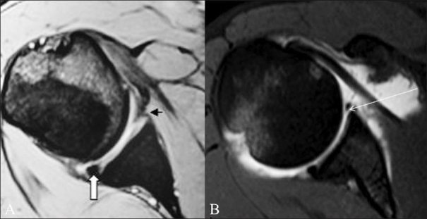
Figure 1 (A,B).

Classic Bankart lesion. A 36-year-old male presented with recurrent anterior shoulder dislocation. Conventional axial GRE MEDIC T2W MRI image (A) shows an attenuated anteroinferior labrum (small arrow). The posterior labrum appears intact and is triangular in shape (thick arrow). TSE T1W fat-saturated axial MRA image (B) shows a classic Bankart lesion as intercalation of contrast material (long arrow) beneath the hypointense anteroinferior labrum
