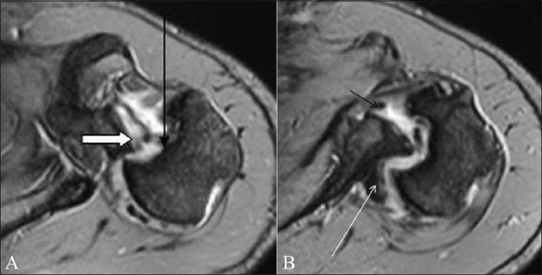
Figure 14 (A-B).

Reverse Hill-Sachs and reverse Bankart lesion in a 34-year-old male who had multidirectional instability with a posterior dislocation at presentation. Axial TSE T1W fat-saturated MRA images (A,B) show a reverse Hill-Sachs lesion (long arrow) as a bony defect in the anterior humeral head. A posterior labral tear is indicated with a thick arrow. The patient also had an anterior labral tear (small arrow in B) and a Hill-Sachs lesion from previous anterior dislocations. A Bennett lesion is seen as ossification, posteriorly along the scapular neck (long arrow in B)
