Figure 17 (A-B).

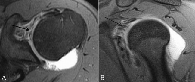
Rotator cuff interval (RCI) tear. Axial TSE T1W fatsaturated MRA image (A) and sagittal TSE T1W fat-saturated MRA image (B) show irregularity of the RCI capsule (arrow). Contrast is seen in the subcoracoid recess

Rotator cuff interval (RCI) tear. Axial TSE T1W fatsaturated MRA image (A) and sagittal TSE T1W fat-saturated MRA image (B) show irregularity of the RCI capsule (arrow). Contrast is seen in the subcoracoid recess