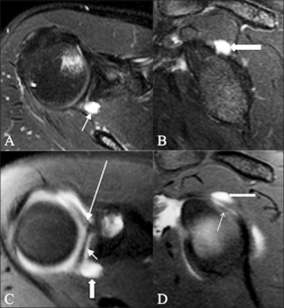
Figure 18 (A-D).

Paralabral cyst. A 22-year-old athlete presented with shoulder pain on overhead throwing movements and instability. TSE T2W fat-saturated axial (A) sagittal (B) MRI images show a welldefined, hyperintense, cystic lesion (arrow) in close relation to the posterosuperior labrum, suggestive of a paralabral cyst. No superior labral pathology is identified. Corresponding axial (C) and sagittal (D) TSE T1W fat-saturated MRA images show a superior labral anteroposterior (SLAP) tear (small arrow); the joint contrast is seen to communicate with the paralabral cyst through the SLAP lesion. The cyst and the insertion of the long head of the biceps tendon are indicated by a thick arrow and a long arrow, respectively
