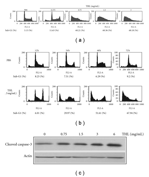
Figure 2.

Effects of THL on the apoptosis induction of NB4 cells. (a) THL induced apoptosis of NB4 cells in a dose-dependent manner after 72 h of treatment. (b) The time course of THL (1.5 mg/mL)-induced apoptosis of NB4 cells. The apoptotic sub-G1 populations in the flow cytometric analysis histograms were obtained as measures of apoptosis and the percentages of them were shown below the histograms. (c) The protein level of cleaved/activated caspase-3 of NB4 cells was increased by THL after 72 h of treatment. The cells were treated with PBS or THL (0.375–3 mg/mL) for 72 h and then harvested for flow cytometry (a and b) and western blot (c) analysis.
