
FIGURE 5.
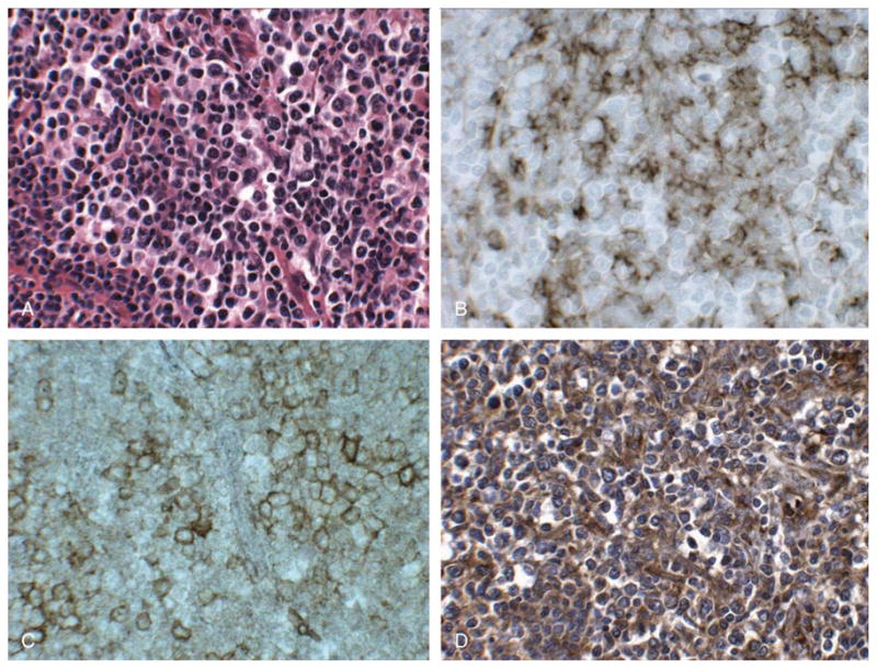
PD-L1 immunostaining in angioimmunoblastic lymphoma. Lymph node involved by angioimmunoblastic lymphoma (A) with CD21-positive FDCs present in expanded networks (B) where foci of PD-1-positive cells are present (C). Immunostaining reveals the presence of numerous PD-L1-positive cells (D) closely associated with PD-1-positive cells. The pattern of PD-L1 staining is similar to that seen with CD21, a marker of FDCs (B). (A, H&E, × 400; B-D, immunostaining with hematoxylin counterstain, × 400).