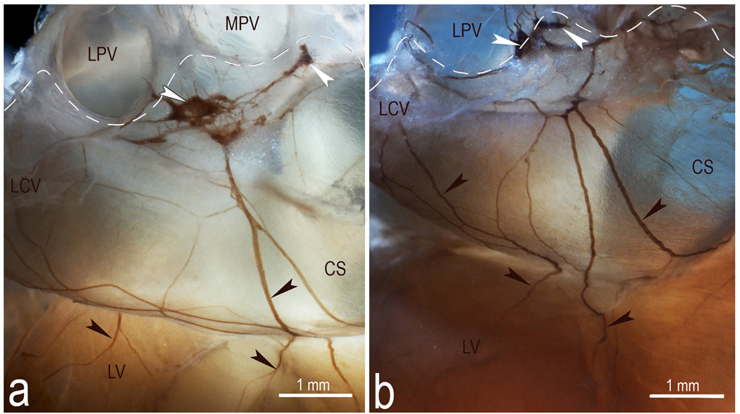
Fig 5.
Macrophotographs illustrating the structural variability of the left dorsal neural subplexus in two mouse hearts stained histochemically for AChE. White arrowheads, some ganglia, black arrowheads, topographically comparable nerves at coronary sinus. Dashed lines demarcate the limits of heart hilum. Abbreviations: CS – coronary sinus; LCV – left cranial (left azygos) vein; LPV – left pulmonary vein; LV – left ventricle; MPV – middle pulmonary vein. Note the persistent location of ganglia specified by white arrowheads.

