
Fig. 2. Persistent LAT clusters caused by over expression of 70Z/3 Cbl contain phosphorylated LAT.
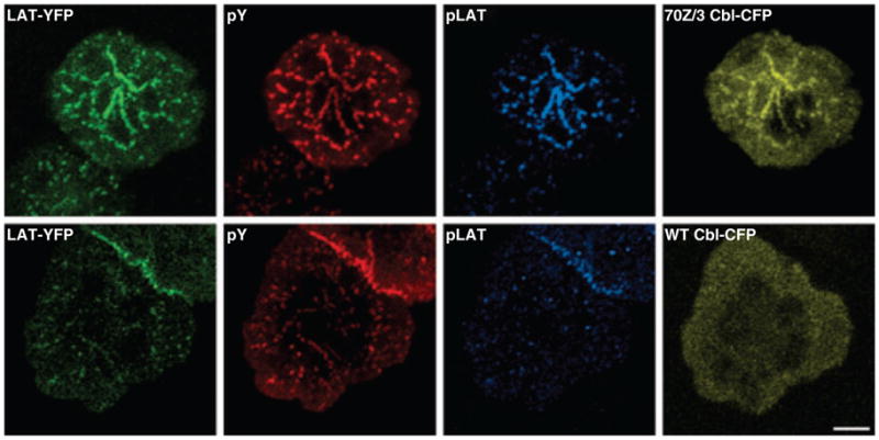
Jurkat T cells expressing LAT–YFP were transfected with either 70Z/3 Cbl–CFP (top row) or wildtype (WT) Cbl–CFP (bottom row). Twenty-four hours after transfection, the cells were plated onto stimulatory coverslips and fixed after incubation at 37 °C for 5 min. The cells were stained with mouse anti-phosphotyrosine (4G10) and rabbit anti-phospho-LAT191. LAT–YFP is shown in the green channel, anti-phosphotyrosine staining in the red channel, anti-phospho-LAT191 staining in cyan, and Cbl–CFP in yellow. In the cells expressing 70Z/3 Cbl-CFP, there are prominent clusters containing both LAT–YFP and 70Z/3 Cbl–CFP that show extensive staining for phosphotyrosine and phospho-LAT. In contrast, cells overexpressing WT Cbl–CFP have faint LAT clusters, no Cbl–CFP clusters, very little phosphotyrosine staining, and almost undetectable phospho-LAT staining. Bar = 5 μm.