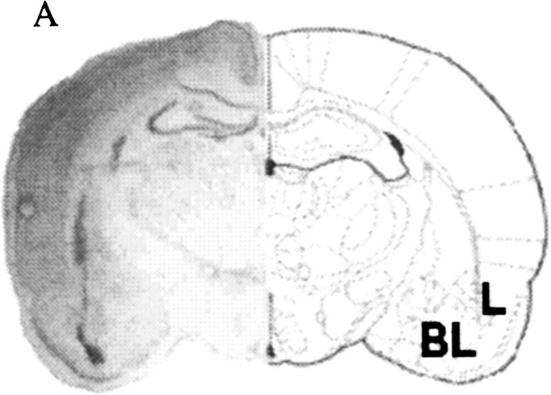
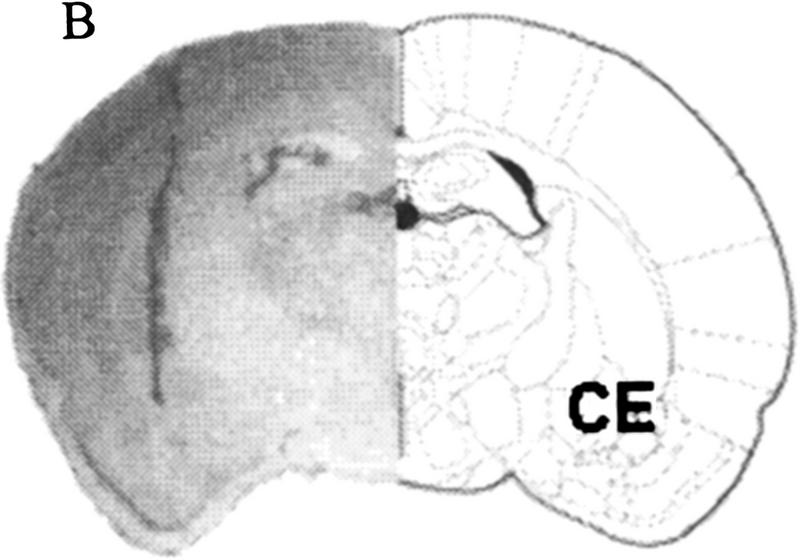
Figure 1.
(A) Localized microinjection of AP5 bilaterally into the lateral/basolateral amygdala. (Left) A photomicrograph of a Nissl-stained section (40 μm) obtained from a rat injected with the competitive NMDA receptor antagonist AP5 (0.5 μl) into the lateral/basolateral nuclei (BL) of the amygdala. (Right) A coronal section from the rat atlas of Paxinos and Watson (1986) that corresponds to the cannula location. (Bregma) −2.56 mm; interaural 6.44 mm. (B) Localized microinjection of AP5 bilaterally into the central amygdala. (Left) A photomicrograph of a Nissl-stained section (40 μm) obtained from a rat injected with the competitive NMDA receptor antagonist AP5 (0.5 μl) into the central nucleus (CE) of the amygdala. (Right) A coronal section from the rat atlas of Paxinos and Watson (1986) that corresponds to the cannula location. (Bregma) −1.80 mm; interaural 7.20 mm.


