
Fig. 2. Expression of Cx36 in trigeminal ganglion neurons is increased in response to CFA.
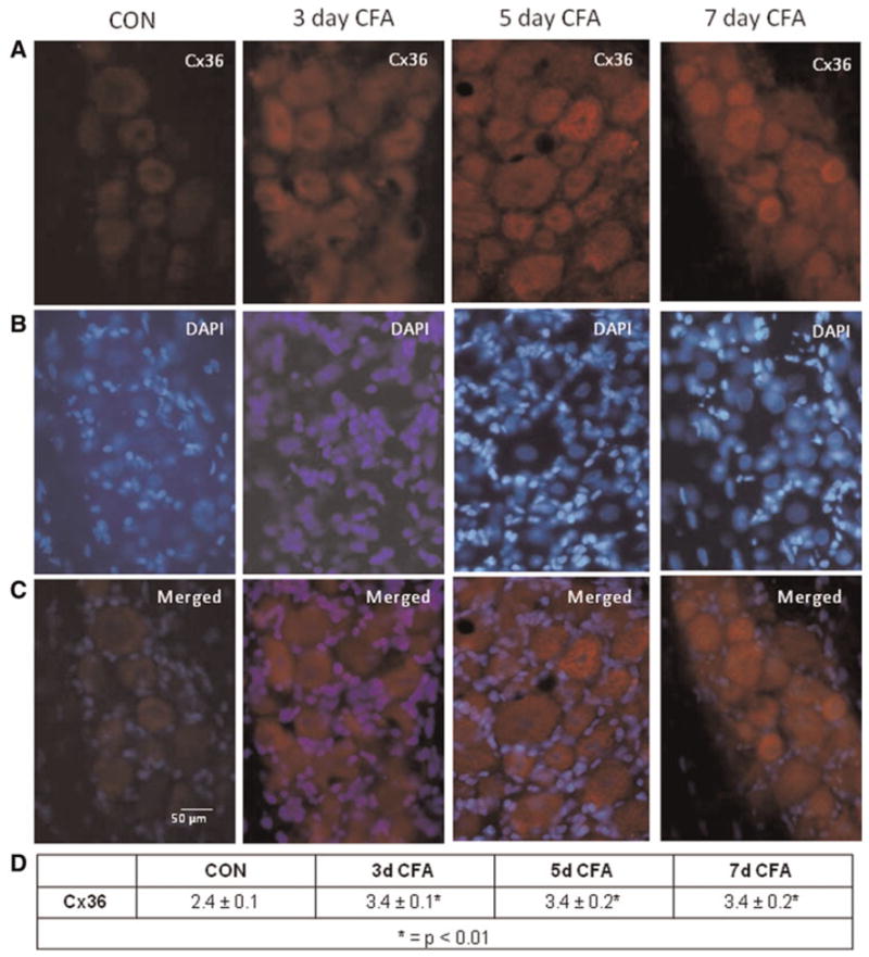
(A) Images (400×) of a representative area in the V3 region of ganglia obtained from control animals (CON) as well as 3, 5 and 7 days after CFA injection in the TMJ capsule are shown. (B) The same tissue sections shown in panel A were costained with the nuclear dye DAPI. (C) A merged image of panels A and B is shown. (D) The average relative staining levels for Cx36 are reported. The normalized values are reported as the average staining intensity ± SEM (n = 3).