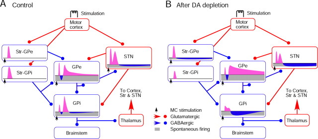
Figure 7.
A, B, Diagrams show major synaptic connections from MC to GPi and a summary of observations from the present study. The connections are based on the data obtained in previous studies (for review, see Kita, 1994, 2007). MC provides strong inputs to Str and STN. In the control group, MC stimulation induces a shorter latency excitation in STN compared with Str due to the unique membrane properties of STN neurons (Farries et al., 2010). The early excitation in STN evokes an early excitation in GPe and GPi before the Str-mediated inhibition begins. The MC-induced excitation in Str neurons projecting to GPi has a longer duration than that in Str neurons projecting to GPe under normal conditions (Flores-Barrera et al., 2010). The late excitation in STN, which may be formed by various forces, drives the late excitation in GPe and GPi. B, In 6-OHDA rats, MC-induced excitation in Str neurons projecting to GPi decreased (Flores-Barrera et al., 2010). Also, spontaneous firing of Str neurons projecting to GPe increased, which induced a tonic inhibition and disinhibition in GPe neurons. The mean latency of the excitation evoked in those neurons was significantly shorter than in the control group and, as a consequence, the early excitation in GPe was reduced. These changes produce a cascade of changes in GPe and GPi. The short inhibition in GPi was almost abolished after DA depletion, and the mechanisms driving this change are still under investigation.

