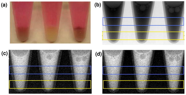
Figure 4.
Pellets of approximately 107 FOCUS cells labeled with 50 nm gold nanoparticles (left), no gold (middle), and 10 nm gold nanoparticles (right). Blue and yellow boxes indicate areas selected for intensity profiles of the supernatant and pellet, respectively. (a) Photograph of pellets under cell culture medium clearly shows gold labeling. (b) X-ray absorption image. (c) Left 1st-order processed image. (d) Upper 1st-order processed image.

